

Rapid Detection of Microorganisms in Traditional Chinese Medicines Using Ultraviolet-Visible Spectro
Introduction
The 2025 edition of the Pharmacopoeia of the People's Republic of China (hereinafter referred to as the Chinese Pharmacopoeia) stipulates explicit microbial limits for multiple Chinese herbal preparations. Consequently, effective detection techniques are required to accurately assess microbial contamination in these preparations, thereby ensuring drug safety. Microorganisms are minuscule and invisible to the naked eye. Traditional detection methods suffer from drawbacks such as high costs and lengthy monitoring times, rendering them unsuitable for large-scale testing requirements. The ultraviolet-visible spectrophotometric method based on silver nanoparticle (AgNP) culture medium primarily employs AgNP medium for microbial cultivation. It measures the absorbance of substances within the 190–800 nm wavelength range, thereby fulfilling requirements for quantitative detection, impurity analysis, and identification. The AgNP-based UV-Vis method offers straightforward operation and high sensitivity. Culturing microorganisms in AgNP medium mitigates various influencing factors, ensuring detection accuracy. UV-Vis light directly interacts with microorganisms, with wavelengths between 200–400 nm sensitively detecting internal nucleic acids and other substances. Analysing spectral information enables microbial species identification. The currently applied UV-Vis spectrophotometry faces limitations in detecting complex backgrounds. Given the intricate composition of traditional Chinese medicines, one-dimensional spectral detection alone struggles to achieve optimal results. Consequently, this study innovates the AgNPs-based UV-Vis spectrophotometry by employing two-dimensional correlation spectroscopy. This transforms one-dimensional spectral digital signals into two-dimensional spectral digital signals, significantly enhancing overall spectral resolution and improving the sensitivity and accuracy of detection outcomes, thereby enabling the identification of microbial species in traditional Chinese medicines. This study selected samples of traditional Chinese medicines such as mulberry mistletoe and two-headed tip to analyse the specific operational procedures and detection results of this method, investigating its value for rapid, large-scale detection of microbial contamination in traditional Chinese medicines.
1 Materials and Methods
1.1 Materials and Reagents
1.1.1 Samples
Eleven batches of two-pointed mulberry mistletoe (Cynomorium songaricum) were selected, originating from Shaanxi, Shandong, Jiangsu, Heilongjiang, Jilin, and Hubei. Twelve batches of mulberry mistletoe (Cynomorium songaricum) were sourced from Anhui, Jilin, Guangdong, and Guangxi. All samples were stored in a designated cool room for later use.
1.1.2 Microbial Strains
Staphylococcus aureus (CMCC(B)26003), Escherichia coli (CMCC(B)44102), Pseudomonas aeruginosa (CMCC(B)10104), Salmonella paratyphi B (CMCC(B)50094), Candida albicans (CMCC(B)98001), Streptococcus pyogenes (CMCC(B)32210), Shigella flexneri (CMCC(B)51572). CMCC denotes the China Medical Culture Collection Centre. Prior to use, prepare bacterial solutions with a colony count below 100 CFU/mL in accordance with the requirements of the Chinese Pharmacopoeia.
1.1.3 Culture Media
The selected culture media comprise AgNPs medium, Columbia CAN blood agar medium, tryptose soy agar (TSA), Sabouraud dextrose agar (SDA), tryptic soy broth (TSB), modified tryptic soy broth, RV Salmonella enrichment broth, pH 7.0 sterile sodium chloride peptone buffer, Kang Kai liquid medium, triple sugar iron agar (TSI), purple bile salt glucose agar, mannitol sodium chloride agar, Bromhexadecyl Trimethylammonium Agar, Enteric Bacteria Enrichment Broth, Broth Medium, MacConkey Agar, Salmon Glucose Broth, Xylose Lysine Deoxycholate Agar, Columbia Blood Agar. All media were supplied as dry powder formulations.
1.1.4 Experimental Instruments and Parameters
Ultraviolet-visible spectrophotometer, electronic balance (accuracy 0.001g), pipette (accuracy ±0.8μL), electric heating constant-temperature incubator (temperature deviation ±0.9℃).
1.2 Experimental Methods
1.2.1 Preparation of Test Solution
Take 25g of the herbal medicine sample, add 225mL of sterile peptone buffer solution at pH 7.0, shake and agitate for approximately 10 minutes, and obtain a test solution with a supernatant-to-solute ratio of 1:10.
1.2.2 Microbial Activation and Isolation
Perform selective microbial culture using AgNPs medium. Place the medium solutions and blank Petri dishes in an autoclave set to 121°C, with pressure at 103.4 kPa and sterilisation time between 15 and 30 minutes. Following sterilisation, place the blank Petri dishes on a sterile bench surface. Cool the sterilised medium to approximately 50°C (to avoid killing microorganisms through excessive heat), pour into the dishes to solidify into plates, and add the test solution heated to 37°C to the dishes. Following these steps, inoculate the bacterial strain into the recovery solution. After a 5-minute interval, inoculate the bacterial suspension into the petri dishes using the streak plate method. Incubate the dishes in a 37°C constant-temperature incubator for 18 hours.
1.2.3 Microbial Propagation and Purification
Weigh LB broth and add liquid at a ratio of 10 g LB broth to 800 mL distilled water. Sterilise via high-pressure steam autoclaving [temperature (115–126°C), pressure (67–139 kPa)]. Allow to cool to 37°C before use. Select individual, independently growing colonies for propagation. Staphylococcus aureus colonies on AgNPs medium appear golden yellow, circular, raised, and smooth-surfaced; Escherichia coli colonies are greyish-white, circular, with neat edges and a moist surface; yeast colonies are milky white. Transfer a single colony to the prepared medium, mix thoroughly, and incubate at 37°C for 18 hours. Following incubation, the separated and propagated bacterial suspension was transferred into centrifuge tubes. Centrifugation (3000 rpm for 5 minutes) was performed, followed by washing with deionised water and removal of the supernatant. The mixture was then resuspended by gentle shaking and centrifuged again (5000 rpm for 10 minutes) to obtain the bacterial suspension.
1.2.4 McFarland Turbidity Method
Prepare McFarland turbidity standard tubes (No. 0.5) with a reference standard of 1.5 × 10⁸ CFU/mL. where 1% sulphuric acid (H₂SO₄) reacts with 1.175% barium chloride (BaCl₂·2H₂O) to form barium sulphate precipitate, creating a stable suspension. Using a pipette, transfer 0.05 mL of the barium sulphate solution (effective working concentration 0.015%) into the Mead's turbidimetric standard tube. Under aseptic conditions, transfer an appropriate volume of microbial suspension into a sterile test tube. Add deionised water to achieve a final microbial suspension concentration of 10⁸ CFU/mL.
1.2.5 Collection of Spectral Data
Add the prepared microbial suspension to the pre-formulated test solution at a ratio of 1 mL test solution to 1mL bacterial suspension, achieving concentrations of 5%, 10%, 15%, 20%, 25%, 30%, and 35%. Following this, add deionised water to bring the total liquid volume to 21mL. Three replicates were prepared for each concentration gradient, yielding a total of 63 samples (7×3×3). The UV-visible spectrophotometer was activated with a preheating temperature of 5–35°C and a preheating duration of 20–30 minutes. Once the light source energy supply stabilised, spectral data collection was conducted according to protocol. Prior to measuring standard samples, pure water was added to seven cuvettes to serve as blank samples. Subsequently, the instrument baseline was calibrated using the front and rear liquid cells. During measurement, ensure the control group's blank samples remain unchanged. Employ spectral scanning mode to sequentially replace samples in the test cell with different concentration gradients, setting sampling intervals to 0.5nm. Thoroughly shake samples before measurement. For each concentration, measure three parallel samples once and calculate the average value. Scan wavelength range: 400–800nm.
1.2.6 Raw Spectral Data and Characteristic Analysis
Following completion of the detection, the UV spectra of traditional Chinese medicine microorganisms were collected, yielding a total of 189 sets of raw data, each comprising 601 wavelength absorbance values. Taking Staphylococcus aureus, Escherichia coli, and yeast as examples: - Staphylococcus aureus exhibited poor absorption characteristics in the 200–300 nm range after addition to the herbal test solution, with multiple absorption peaks emerging beyond 300 nm; the first peak occurred around 334 nm. Escherichia coli exhibited poor absorption characteristics in the 200–300 nm range when added to the herbal test solution, producing multiple absorption peaks beyond 300 nm. with the primary absorption peak occurring around 331 nm. Furthermore, the presence of light-absorbing substances such as fat globules and casein gel particles in the herbal test solution results in a generally elevated overall absorbance. The primary light-absorbing components of E. coli in the UV-visible spectrum include tryptophan, phenylalanine, and tyrosine, with absorption wavelengths at 279 nm, 259 nm, and 278 nm respectively. Following the addition of yeast to the herbal test solution, absorption characteristics within the 200–300 nm range were suboptimal, with multiple absorption peaks emerging beyond 300 nm. The spectral curve exhibited in the 300–350 nm range did not display a uniform upward trend, primarily due to the uneven distribution and varying sizes of the yeast cells. Observation revealed the first absorption peak at 337 nm, with overlapping light-absorbing substances between different components.
Results and Analysis
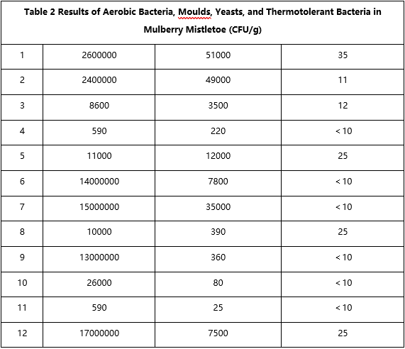
Eleven batches of Dendrobium officinale and twelve batches of Taxillus chinensis exhibited severe microbial contamination. Among these, Dendrobium officinale recorded maximum aerobic bacteria counts of 1,600,000 CFU/g, maximum mould and yeast counts of 1,200 CFU/g, and maximum thermotolerant bacteria counts of 18 CFU/g. For mulberry mistletoe, the highest aerobic bacteria count reached 21,000,000 CFU/g, with mould and yeast at 51,000 CFU/g and thermotolerant bacteria at 35 CFU/g. See Tables 1 and 2.

3 Discussion and Conclusions
To ensure the safety and efficacy of Chinese herbal medicines, authoritative publications such as the Chinese Pharmacopoeia stipulate explicit microbial limits for herbal preparations, thereby advancing the internationalisation and modernisation of traditional Chinese medicine. Consequently, relevant institutions must select the most effective technical protocols for microbial testing during quality control procedures, ensuring that microbial counts in marketed herbal preparations remain within acceptable ranges [5].
Data from this study indicate that 11 batches of Dipterocarpus retusus and 12 batches of Taxillus chinensis exhibited severe microbial contamination, with detectable levels of aerobic bacteria, moulds, yeasts, and thermotolerant bacteria. Control microorganism testing revealed that 11 batches of Lianhuasheng and 12 batches of Sangjisheng contained special types of microorganisms, bile salt-tolerant Gram-negative bacteria, and typical bacteria. These findings align with the conclusions of Hu Yuxia et al., suggesting that the ultraviolet-visible spectrophotometric method based on AgNPs medium enables rapid and accurate detection of microbial content in traditional Chinese medicines. Analysis of the results indicates that the predominant types of thermotolerant bacteria are spore-forming bacteria and spores, characterised by strong heat resistance and difficulty in eradication. While the Pharmacopoeia does not specify an upper limit for thermotolerant bacteria, it is important to recognise their potential hazards as they cannot be completely eliminated even after decoction. Effective measures must therefore be taken to ensure their thorough removal. Bile salt-tolerant Gram-negative bacteria, belonging to the non-lactose fermenting group, exhibit tolerance to bile salts and efficient utilisation of glucose. The Pharmacopoeia has incorporated these into the mandatory microbial testing scope. Significant variations exist in the contamination levels of aerobic bacteria, moulds, and yeasts across different types of Chinese herbal decoction pieces. As the Pharmacopoeia does not provide uniform regulations, relevant enterprises may determine specific control schemes based on actual circumstances. The foregoing analysis demonstrates that the UV-visible spectrophotometric method utilising AgNPs-based media enables rapid detection of microbial contamination in traditional Chinese medicines. However, it demands a high level of technical proficiency from operators. Consequently, during application of this technique, factors influencing results must be clearly identified, and the testing protocol optimised according to practical conditions. Spectral data acquisition requires multiple measurements with averaging, alongside mastery of the spectral characteristics of various microorganisms to ensure detection accuracy.
In summary, the UV-visible spectroscopy method utilising AgNPs-based culture medium offers rapid and precise detection of microbial contamination in traditional Chinese medicines, demonstrating significant potential for practical application. However, the study's duration was relatively brief, and the sample size of herbal medicine preparations was insufficient. Future microbial testing of traditional Chinese medicines should therefore focus on refining the testing protocol, controlling factors influencing results, and standardising operational procedures to guarantee the accuracy of findings.
